Blood from the heart is mainly ejected upwards along the ascending aorta. When pulling blood into the heart, the major motion is also along the axis parallel to the spine. Thus the major motion is longitudinal. For both ejecting and pulling blood, according to Newton's 3rd Law the force exerted on the blood by the heart is matched by an equal and opposite force on the body by the blood.
If a patient is placed on a table with very low friction, then the force on the body causes the body and the table to move back and forth as the blood is being pumped. A sensitive accelerometer on the table measures its acceleration, and one can compute the acceleration of the blood with: This is called a ballistocardiogram (BCG), and an apparatus to make one is shown to the right. The apparatus was made by Nihon Kohden in 1953, and we use the figure with permission. The original figure is at http://www.nihonkohden.com/50th/history2.html |
 |
Although the technique has been known for over 50 years, because the mass of the accelerating blood is small compared to the mass of the body and table, the experimental errors in the measured blood acceleration were large. Thus it was not very useful as a diagnostic tool. Recently modern signal processing techniques have allowed these experimental errors to be greatly reduced, and BCG's are now often used in a medical context.
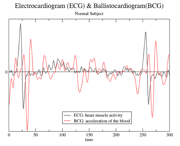
As you will learn in the third quarter, the electrocardiogram (ECG) measures the activity of the muscles in the heart; there is also an experiment in the laboratory on ECGs. The BCG measures the effect of this muscle activity by directly measuring the acceleration of the blood. The following figure shows an ECG and a BCG for a normal patient.
 |
The data for the above figure was supplied to us by Dr. William McKay, Department of Anesthesia, University of Saskatchewan.
This document was written by David M. Harrison, Dept. of Physics, Univ. of Toronto, in July 2003.